Pelvic ultrasound is performed to assess the pelvic structures in patients with various gynaecological complaints. It uses sound waves to produce images of pelvic structures including uterus and ovaries. Similar ultrasound technology is also used in pregnancy to evaluate the baby. Recent advancement is 3-D imaging using a transvaginal or a trans abdominal approach approach. 3-D ultrasound offers an advantage of producing a reconstructed image using the transverse and longitudinal images in gynaecology. The various applications are:
- Sono AVC where the ovarian follicles are monitored and shown
- 3-D UTERUS - where the exact shape of the cavity of the uterus is shown in the coronal plane. This imaging can almost replace MRI and Hysteroscopy.
- 3D VOCAL - Where the volume of the endometrial cavity is calculated and shown as a 3-D image.
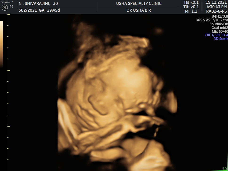
- 3-D obstetrics - The various organs in the baby are better visualised to assess abnormalities.the face of the baby can be visualised in this 3-D model.
Trans abdominal Ultrasound
Your doctor would place an ultrasound probe over the abdomen after the application of ultrasound gel. Your doctor may ask you to keep the bladder full while performing the ultrasound.
Transvaginal Ultrasound
Your doctor would place a thin probe into the vagina after the application of lubricating gel to visualise the uterus and ovaries. please follow the instructions given by your doctor during the procedure. remember to relax yourself and that the procedure is not a painful one.Trans vaginal approach of ultrasound always gives better images of pelvic structures as the ultrasound probe is placed close to the organs of interest.